# Brain Reorganization After Nerve Repair Surgery Offers New Clues for Hand Recovery
Surgeons can now restore physical connection between a severed peripheral nerve and the hand, but the brain does not simply resume its original function. Research reveals the brain reorganizes how it processes signals from individual fingers following nerve repair surgery, a finding that could reshape recovery protocols and rehabilitation strategies.
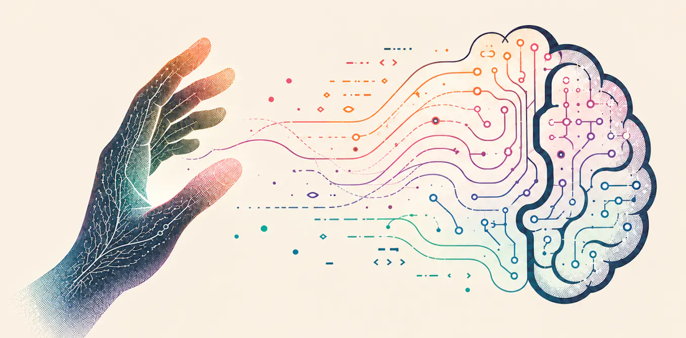
When a peripheral nerve tears, the link between hand and central nervous system breaks. Microsurgical techniques have made nerve repair possible, restoring the anatomical pathway for sensation and motor control. Yet patients rarely regain full function. The new research suggests why. The brain's sensory and motor cortices, which map each finger with precision in healthy people, reconfigure after repair surgery.
During nerve repair recovery, the brain does not simply reactivate dormant finger representations. Instead, the neural tissue reorganizes. Individual fingers may activate larger or overlapping brain regions than before injury. Some fingers might recruit unexpected brain areas. The cortical map becomes less precise.
This neuroplasticity, while allowing some recovery, explains why patients often experience persistent numbness, tingling, or reduced dexterity. The hand sends signals the brain no longer recognizes with its original specificity. Communication exists, but translation becomes imperfect.
The implications extend across rehabilitation practice. Current recovery programs focus primarily on hand exercises and sensory reeducation, assuming the brain will naturally reclaim its previous architecture. If cortical reorganization is permanent or difficult to reverse, rehabilitation strategies need adjustment.
Targeted sensory training might help. By deliberately stimulating individual fingers during recovery, patients could reinforce cortical representations and guide the brain toward more precise mapping. Early intervention proves critical. The brain's reorganization happens quickly after nerve injury, making the first weeks and months after surgery especially important for shaping recovery outcomes.
Physical and occupational therapists could integrate this knowledge into post-surgical protocols. Rather than generic hand exercises, therapy could emphasize discriminative touch tasks, where patients learn to distinguish which finger is being touched without looking. This trains the brain to sharpen its finger-specific processing.
Researchers are also exploring whether visual feedback and mental imagery during rehabilitation help maintain or restore cortical organization. Virtual reality tools and neurofeedback systems might accelerate cortical remapping in ways traditional therapy cannot.
The discovery underscores a broader principle in neuroscience. Injury does not simply break a circuit that surgery can repair. The nervous system actively reconstructs itself after damage. Understanding that reconstruction process opens doors to smarter, more targeted interventions.
Patients who undergo peripheral nerve repair today should expect realistic outcomes based on this emerging science. Full sensation and motor control rarely return completely. But informed rehabilitation, delivered early and targeted toward cortical reorganization, can optimize whatever recovery the brain achieves. The goal shifts from restoring the original brain map to developing the most functional new map the brain can build.
This research should prompt collaboration between surgeons, neuroscientists, and rehabilitation specialists. No single discipline holds all answers to nerve recovery. Coordinated care, informed by evidence about cortical plasticity, offers the best path forward for patients facing the long recovery after nerve repair surgery.